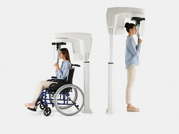
Carestream Panoramic Dental X-Ray

Carestream panoramik röntgen sistemi, dişlerin, çene kemiklerinin ve çevre anatomik yapıların geniş bir alanda görüntülenerek değerlendirilmesini destekler.

What Is the Carestream Panoramic X-Ray System?

The Carestream panoramic X-ray system captures a broad digital radiographic view of the mouth, teeth and jaws.

A panoramic X-ray provides an overview of the upper and lower teeth, jawbones and related anatomical structures in a single image. It supports diagnosis and treatment planning by providing additional information about areas that cannot be seen directly during a clinical examination.

Panoramic imaging can be used for a range of clinical needs, from assessing impacted teeth to reviewing overall oral health. Whether an X-ray is needed is determined individually following a clinical assessment.

Carestream Panoramic Imaging at DentLotus

At DentLotus, we use Carestream panoramic imaging where needed to support the clinical examination and provide a broader assessment of the mouth and jaws.

The digital panoramic image allows us to review tooth positions, jawbones, impacted or unerupted teeth and related anatomical structures together.

Considering radiographic findings alongside the clinical examination helps the dentist assess your condition more fully and plan any treatment needed. Reviewing digital images with you can also help make the treatment process easier to understand.

Key Features of Carestream Panoramic Imaging

A Broad View of the Teeth and Jaws

Allows assessment of the upper and lower teeth and surrounding structures in one panoramic image.

Digital Images

Displays the captured radiograph digitally for the dentist to review.

Comprehensive Assessment

Helps assess dental and jaw structures that cannot be seen directly during examination.

Treatment Planning

Supports treatment planning by bringing together radiographic findings and the clinical examination.

How Panoramic Imaging Supports Your Treatment

A clinical examination is the foundation of assessing your teeth and surrounding tissues. Radiographs may also be needed to examine tooth roots, jawbone, impacted teeth and certain anatomical structures.

A panoramic X-ray provides an overview of the mouth and jaws by capturing a wide area in one image. Considered alongside clinical findings, it can help identify treatment needs.

If a panoramic image does not provide enough detail, targeted imaging such as RVG or three-dimensional imaging may also be used according to clinical needs.

Where Is Panoramic Imaging Used?

Panoramic X-rays can support diagnosis and planning across dental treatments that require an overview of the mouth and jaws.

Dental Implant Treatment

Can help assess the overall condition of the teeth and jawbone before implant treatment. Three-dimensional imaging may also be needed for detailed implant planning.

Orthodontics

Panoramic images can help assess tooth development, eruption and overall positions within the jaws.

Oral and Maxillofacial Surgery

Can provide an overview of the positions of impacted or unerupted teeth and their relationship to nearby anatomical structures.

Periodontics

Can support a clinical periodontal examination by providing an overview of the bone supporting the teeth.

What Happens During a Panoramic X-Ray?

Following a clinical assessment, you are positioned in the panoramic X-ray unit. The image is then captured and reviewed digitally.

1

Clinical Assessment

The dentist examines your mouth and teeth to determine whether a panoramic X-ray is needed.
2

Getting into Position

You are carefully positioned in the panoramic X-ray unit to help capture an accurate image.
3

Capturing the Panoramic Image

The unit moves around your head in a controlled way to capture a panoramic radiograph of your mouth and jaws.
4

Review and Planning

The dentist reviews the digital image alongside clinical findings and uses it for treatment planning where needed.
Frequently Asked Questions

Your Panoramic X-Ray Questions Answered

What Is a Panoramic Dental X-Ray?

A panoramic dental X-ray captures the upper and lower teeth, jawbones and certain surrounding anatomical structures in one broad image.

What Can a Panoramic X-Ray Show?

It can help assess overall tooth positions, unerupted or impacted teeth, jawbones and certain surrounding structures. The image is interpreted alongside the clinical examination.

How Does a Panoramic X-Ray Differ from RVG?

A panoramic X-ray provides a broad view of the mouth and jaws. RVG is used for a detailed radiographic assessment of one tooth or a smaller area.

Is a Panoramic X-Ray Painful?

No. Imaging involves no surgical or invasive procedure inside the mouth. You simply need to remain still briefly in the correct position.

Does a Panoramic X-Ray Use Radiation?

Yes. Panoramic images are created using X-rays. Your dentist therefore assesses whether imaging is needed according to clinical requirements.

Do I Need a Panoramic X-Ray Before Every Dental Treatment?

No. A panoramic X-ray is not needed for every patient or treatment. The imaging required depends on examination findings and the planned treatment.
Our Treatments

Treatments Supported by Panoramic Imaging

Panoramic imaging can support diagnosis and planning for treatments that require an overview of the mouth and jaws.

Periodontology

  • Diş Eti Hastalıkları
  • Diş Eti Kanaması
  • Diş Eti Çekilmesi
  • Periodontal Tedavi
Explore This Treatment
01

Periodontology

Orthodontics

  • Diş Teli Tedavisi
  • Şeffaf Plaklar
  • Kapanış Bozuklukları
  • Ortodontik Takip
Explore This Treatment
02

Orthodontics

Dental Implants

  • Eksik Diş Tedavisi
  • İmplant Planlaması
  • İmplant Uygulaması
  • İmplant Üstü Protez
Explore This Treatment
03

Dental Implants

Explore Your Treatment Options